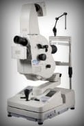
As the old adage goes, “The eyes are the window of the soul”, the state of the mind of a person can be very easily ascertained by looking at the emotions exhibited by a person’s eye. The eyes of humans are beyond compare and much more sophisticated than even the images which are produced by the best supercomputers and no other species on this earth has visual sense organs like humans. The human eyes can sense a wide range of colours and images. Therefore if anything happens to the eyes, the person is devastated. There are many diseases such as Diabetes which exert a bad influence on the eyes and the person slowly loses sight. Fortunately most of the incidence of blindness can be prevented easily with proper diagnostics with special optical equipments. One such ophthalmic equipment which is used for detecting visual problems is the fundus camera.

Retinal camera or the fundus camera is an important diagnosing tool for eye ailments. The retina is the most important part of the eye since it is the place where the image is formed and the image is transmitted to the brain for analysis and image formation. The retina is much akin to the photographic plate in any camera. A retinal camera is a camera with a microscope and an eyepiece for viewing the image of the retina. Fundus camera or the retinal camera can also photograph the image of the retina.
A Fundus camera unlike an ordinary camera which gives about 50 degrees of the retinal area and a 2.5X magnification provides a 140 degree view of the retina and 5X magnification. The retinal camera uses a system of lenses through which light is focused on the retina after passing through the cornea. The light is then reflected by the retina and traces a different path to the eyepiece. Since the paths of the incident and the reflected light are different, there is no risk of the two mixing up and causing any distortion.
Ophthalmic equipment like the retinal camera finds a number of applications. The camera is used to take colour pictures of the retina for studying any anomaly in the structure. The camera is also equipped to filter off the red colour so that the blood vessels are more clearly visible since the blood vessel will appear dark in any other colour except red colour. The camera can also be used for retinal angiography. A florescent or contrasting medium is introduced in the blood vessel so that the fine vessels in the retina are visible and any blockages can be seen clearly.
